.png)

ECG ANALYSIS
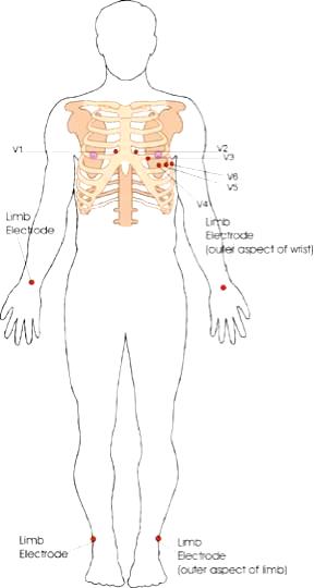
The 12-lead ECG misleadingly only has 10 electrodes
(sometimes also called leads but to avoid confusion we will refer to them as electrodes).
The leads can be thought of as taking a picture of the heart’s electrical activity
from 12 different positions
using information picked up by the 10 electrodes.
These comprise 4 limb electrodes and 6 chest electrodes.
When electrical activity (or depolarisation)
travels towards a lead, the deflection is net positive.
When the activity travels away from the lead
the deflection is net negative.
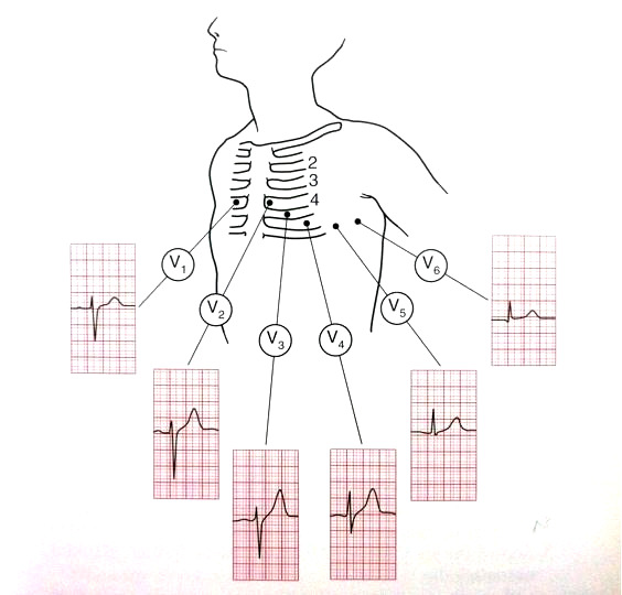
If it is at 90 degrees then the complex is ‘isoelectric’
i.e. the R and S wave are the same size.
This can often be seen in V4 (see Figure 3).
travels towards a lead, the deflection is net positive.
When the activity travels away from the lead
the deflection is net negative.
If it is at 90 degrees then the complex is ‘isoelectric’
i.e. the R and S wave are the same size.
This can often be seen in V4 (see Figure 3).
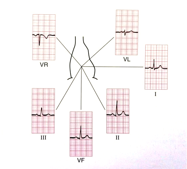
THE LIMB LEADS OR ELECTRICAL VIEWS
THE CHEST LEADS OR ELECTRICAL VIEWS
The areas represented on the ECG are summarized below:
THE 90 DEGREE ANGLE
Lead I = L side of the heart
aVF = inferior territory (remember ‘F’ for ‘feet’)
THE DIAGNOALS
aVR = R side of the heart aVL = L side of the heart
Lead III = inferior territory Lead II = inferior territory
THE 90 DEGREE ANGLE
Lead I = L side of the heart
aVF = inferior territory (remember ‘F’ for ‘feet’)
THE DIAGNOALS
aVR = R side of the heart aVL = L side of the heart
Lead III = inferior territory Lead II = inferior territory
The areas represented on the ECG are summarized below:
V1, V2 = RV V3, V4 = septum V5, V6 = L side of the heart
V1, V2 = RV V3, V4 = septum V5, V6 = L side of the heart