.png)

ECG ANALYSIS
Introduction
Conduction through the heart is dependent on pacemaker cells,
which are organised into key structures.
The heart is a dual pump that sits at the centre of the cardiovascular system.
It is composed of both contractile cells and autorhythmic cells
(also known as pacemaker cells).
Approximately 1% of cardiac tissue is composed pacemaker cells,
which are organised into key structures
and can undergo spontaneous depolarisation.
Depolarisation refers to the electrical changes that occur within a muscle to allow it to contract.
The heart is essentially one big muscle that can contract by itself.
We can detect these electrical changes,
which are associated with heart muscle contraction,
using an ECG.
Conduction system
The sinoatrial node initiates the electrical impulses
that stimulate muscle contraction.
The hearts pacemaker cells are organised into structures including
The Sinoatrial node (SAN),
The Atrioventricular node (AVN),
The Bundle of His,
The right bundle branches
The left bundle branches
The Purkinje fibres.
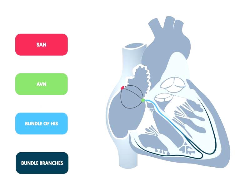
Structural organisation of the hearts pacemaker cells
means there is coordinated electrical activity,
which leads to a coordinated contraction of both atria and ventricles.
Each structure has an intrinsic rate of autorhythmicity.
This refers to the number of electrical signals, and thus contractions, that will occur each minute.
Electrical activity starts with the SAN,
which has the highest rate of autorhythmicity at 70-100 bpm.
Depolarisations of the SAN spread throughout the atria
and reaches the AVN.
Electrical activity then travels through the bundle of His and bundle branches.
Finally the Electrical activity spreads at a slower pace along the purkinje fibres and into the ventricular muscle, leading to coordinated ventricular contraction.